Introduction:
Human thoughts, feelings, and
actions begin in the central nervous system. The brain acts as the primary
mediator- organ, controlling and determining how people interact with the
world. All human responses are the result of the complex interaction between
underlying neuroanatomy and neurophysiology, as well as genetic, environmental
and development factors.
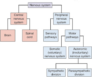
The nervous system is your body's
decision and communication center. The central nervous system (CNS) is made of
the brain and the spinal cord and the peripheral nervous system (PNS) is made
of nerves. Together they control every part of your daily life, from breathing
and blinking to helping you memorize facts for a test. Nerves reach from your
brain to your face, ears, eyes, nose, and spinal cord... and from the spinal
cord to the rest of your body. Sensory nerves gather information from the
environment; send that information to the spinal cord, which then speed the
message to the brain. The brain then makes sense of that message and fires offa
response. Motor neurons deliver the instructions from the brain to the rest of
your body. The spinal cord, made of a bundle of nerves running up and down the
spine, is similar to a superhighway, speeding messages to and from the brain at
every second.
Brain
·
Brain is the most important structure in the
human body. Weighs 3 to 5 pounds, the brain contains approximately 140 billion
cells, making it the most complicated and vital organ in the body. Brain cells
are categorized as either neurons or neuroglia. Neurons generate and
conduct electrical signals.
·
Neuroglia provides the mechanical and
physiological support to the neurons.
·
White matter is composed of the axons of neurons that are insulated by myelin. White matter
makes up the core of major brain structures.
·
The gray matter or cortex typically
covers the surface of these organs.
·
The cortex is functional area of the brain where
neurons communicate with each other and where neurotransmitters are
concentrated.
Parts of the brain
·
The brain is made of three main parts: the
forebrain, midbrain, and hindbrain.
·
The forebrain consists of the cerebrum and
diencephalon(thalamus, hypothalamus and limbic system).
·
The midbrain consists of the mesencephalon
(tectum and tegmentum).
·
The hindbrain is made of the cerebellum, pons
and medulla. Often the midbrain, pons, and medulla are referred to together as
the brainstem.
The Cerebrum: The cerebrum or
cortex is the largest part of the human brain, associated with higher brain
ftlnction such as thought and action. The cerebral cortex is divided into
four sections, called "lobes": the frontal lobe, parietal lobe,
occipital lobe, and temporal lobe.
Functions of these lobes-
·
Frontal Lobe- associated with reasoning,
planning, parts of speech, movement, emotions, and problem solving
·
Parietal Lobe- associated with movement,
orientation, recognition, perception of stimuli
·
Occipital Lobe- associated with visual
processing
·
Temporal Lobe- associated with perception and
recognition of auditory stimuli, memory, and speech
Normal functions and symptoms of
dysfunction of the cerebrum:
|
Lobe
|
Location
|
Normal function
|
Symptoms of alteration
|
|
Frontal
|
Anterior or front area of the brain
|
Programming and execution of motor functions, higher thought process,
intellectual insight, judgment, expression of emotion, decision making.
|
Changes in affect, alteration in language production, motor functioning,
decision making, impulsive behavior
|
|
Parietal
|
Posterior to
central sulcus
|
Sensory
perception,following directions on a map, reading a clock, or dressing
oneself.
|
Decreased
consciousness of pain sensation, difficulty with time concepts, alteration in
personal hygiene, inability to calculate numbers or perform motor actions,
poor attention span.
|
|
Temporal
|
Lies beneath skull on both sides
|
Primarily responsible for hearing and receiving information via ears
|
Auditory hallucinations, increased sexual focus, decreased motivation,
alterations in memory and emotional responses, sensory aphasia
|
|
Occipital
|
Most posterior of
brain
|
Seeing and
receiving information via eyes
|
Visual
hallucinations
|
The cerebral cortex is highly
wrinkled. Essentially this makes the brain more efficient, because it can
increase the surface area of the brain and the amount of neurons within it.
A deep furrow divides the cerebrum into two
halves, known as the left and right hemispheres. The two hemispheres look
mostly symmetrical yet it has been shown that each side functions slightly
different than the other. Sometimes the right hemisphere is associated with
creativity and the left hemisphere is associated with logic abilities. The
corpus callosum is a bundle of axons which connects these two hemispheres.
Nerve cells make up the gray surface
of the cerebrum which is a little thicker than your thumb. White nerve fibers
underneath carry signals between the nerve cells and other parts of the brain
and body.
Diencephalon: Constitutes
only 2% of the CNS by weight.Has extremely wide spread and important
connections; the great majority of sensory, motor and limbic pathways involve
the diencephalon. It includes: thalamus, hypothalamus and the limbic system.
Mesencephalon: Structures of
major importance of mesencephalon or midbrain include nuclei, and fiber tracts.
It extend from the pons to the hypothalamus and is responsible for integration
of various reflexes, including visual, auditory and righting reflexes.
Pons: Is a bulbous structure
that lies between the mid brain and the medulla. Composed of large bundles of
fibers and forms a major connection between the cerebellum and the brain stem.
Also contains the central connections of cranial nerve V through VIII and
center for respiration and skeletal muscle tone.
Medulla: Is the connecting
structure between the spinal cord and the pons, and all of the ascending and
descending fiber tracts pass through it. The vital centers are contained within
the medulla , and is responsible for regulation of heart rate, blood pressure
and respiration. Reflex centers for swallowing, sneezing, coughing and
vomiting.
Basal nuclei:
Also known as basal ganglia are
concentrations of cell bodies closely involved in motor functions and
association. They are concentrations of grey matter located within the white
matter of the cerebrum and midbrain. Among the most well known basal nuclei are
caudate lobe, putamen, globus pallidus, and substantia nigra. Functions
of these basal nuclei are:
·
Translate rnovements such as walking while it is
happening, and they also modulate and correct muscle functioning that allows
movement to occur in a coordinated manner.
·
Aid in the learning and programming of the
complex motor behavior which in the course of time becomes automatic.
Conditions such as Huntington's
disease and Parkinson's disease are associated with basal nuclear dysfunction
and their inability to effectively communicate with the cerebral cortex. Even
some medications given to treat psychiatry disorders alter the basal nuclei,
example: chlorpromazine and haloperidol are two older neuroleptic antipsychotic
medications that cause hypertonicity or dystonia.
Some of the network of nuclei and
their functions:
l . Cerebral cortex: involved in
critical decision making and higher order thinking such as abstract reasoning.
2. Limbic system: involved in
regulating emotional behavior, memory and learning.
3. Basal ganglia: co-ordinates
involuntary movements and muscle tone.
4. Hypothalamus: regulates pituitary
homones.
5. Locus ceruleus: makes norepinephrine,
a neurotransmitter involved in response to stress. It's located in the pons
region of the brain.
6. Raphe nuclei: makes serotonin,
involved in regulation of sleep and behavior mood.
The Cerebellum
The cerebellum, or "little
brain", is similar to the cerebrum in that it has two hemispheres and has
a highly folded surface or cortex. This structure is associated with regulation
and coordination of movement, posture, and balance. It is situated at the base
of the skull, above the brainstem and beneath the occipital lobe of the
cerebral cortex.
Functions:
·
Fine Movement Coordination
·
Balance and Equilibrium
·
Muscle Tone
Limbic system:
Instincts, primitive drives, sexual
arousal, fear, aggression and other emotions are part of the functions of the
structures deep within the brain called the limbic system or limbic lobe. The
structures of this system are:
-- Hippocampus
-- Thalamus
-- Hypothalamus
-- Amygdala
-- Limbic Midbrain Nuclei
Hippocampus
·
Involved in storing information, especially the emotions
attached to memory.
·
Deterioration of these nerves lead to hallmark
symptoms of memory dysfunction.
·
Damage to the left hippocampus impairs verbal memory and damage to the right one causes difficulty with recognition and recall of
complex visual and auditory
patterns.
·
Modulates emotional states,
·
Regulates affective responses to events
·
Difficulties in memory of Alzheimer disorder.
Thalamus
·
Relay switching center of the brain
·
Thus it prevents the cortex from becoming
overload with sensory stimulus.
·
Damage to a very small area will cause defect in
many cortical functions, thus causing behavioral problems.
·
Influences prefrontal cortical functions such as
affect and foresight.
·
influences mood and general body movements
associated with strong emotions, such as fear or rage.
Hypothalamus
·
Basic human activities such as sleep-rest patterns, body temperature, thirst, and
physical drives(hunger and sex) are regulated by this area.
·
Situated deep within the brain.
·
Dysfunction of this structure causes sleep and
appetite problems.
·
Also helps in the production of ADH, and
Oxytocin.
·
Research indicates that some symptomatic
behaviours, such as appetite and sleep problems in the depressed clients, the
seasonal mood changes of SAD (Seasonal Affective disorder), and temperature regulation
problems in clients with schizophrenia.
Amygdala
·
This is directly connected to more primitive
centers of the brain. Lies adjacent to
the hippocampus
·
This provides an emotional component to memory
and is involved in modulating sexuality and aggression.
·
Impulsive acts of aggression and violence have
been linked to deregulation.
·
Modulates emotional states, regulates affective
responses to events.
·
Rapid misfiring of neurons in this regions
causes typical symptoms of BPAD and panic disorder.
Limbic midbrain nuclei
·
This is a collection of neurons that appear to
play a role in the biologic basis of addiction.
·
Sometimes referred to as the pleasure or reward
center of the brain.
·
The main function is to reinforce chemically
certain behavior, ensuring their repetition.
|
Structure
|
Function
|
Dysfunction
|
|
Amygdala
|
Modulates emotional states. Regulates affective responses to events
|
Rapid misfiring of neurons in this region causes typical symptoms of
BPAD. panic disorder.
|
|
Thalamus
|
Relays all
sensory information, except smell. Filters incoming information regarding
emotions and memory to prevent cortex overloading.
|
|
|
Hypothalamus
|
Regulates basic human functions such as sleep-rest patterns, body
temperature, and physical drives of hunger and sex.
|
Symptomatic behavior in depressed clients, seasonal mood changes in SAD
and temperature regulation problem in schizophrenic.
|
|
Hippocampus
|
Modulates
emotional states, Regulates affective responses to events
|
Difficulties in
memory of Alzheimer disorder.
|
Brain stem
Brainstem is the posterior part of
the brain, adjoining and structurally continuous with the spinal cord.
The brain stem provides the main
motor and sensory innervation to the face and neck via the cranial nerves. This
is an extremely important part of the brain as the nerve connections of the
motor and sensory systems from the main part of the brain to the rest of the
body pass through the brain stem.
This includes the corticospinal
tract (motor), the posterior column-medial lemniscus pathway (fine touch,
vibration sensation and proprioception) and the spinothalamic tract (pain,
temperature, itch and crude touch). Brain stem regulates the central nervous
system, and is pivotal in maintaining consciousness and regulating the sleep
cycle. The brain stem has many basic functions including heart rate, breathing,
sleeping and eating.
Clinical application
1.Schizophrenia
Researchers have noted that an increase
in ventricular size is apparent in many people with schizophrenia. In
schizophrenia increased ventricle size is most likely related to
neurodevelopmental factors; that is, the brain around the ventricles has failed
to develop, and the ventricles have enlarged to fill the empty face.
Other biologic differences found in
people with schizophrenia include a decrease in cerebral blood flow, particularly
in the prefrontal areas of the cortex. Imaging technologies that tracks blood
flow and glucose metabolism has substantiated this physiologic change. These
brain changes result in a decline in frontal cognitive functions, such as
organizing, planning, learning, problem solving and critical thinking. Another
biological theory (dopamine hypothesis), it is caused by alterations of
dopamine levels in the brain.
2.Depression
Decreased amounts of
norepinephrine and serotonin, two important brain neurotransmitters, are
thought to play a role in depression. Some researchers have found evidence for
the involvement of acetylcholine, dopamine, and GABA. The receptor, thyroid,
hypothalamic, and pituitary has a role in depression.
3.Anxiety
Research has indicated that drugs
that activate GABA receptors, causing an inhibitory effect, can calm anxious
patients. Other neurotransmitters, such as norepinephrine, epinephrine,
dopamine, and serotonin might also have roles in anxiety.
4.Dementias
Alzheimer's disease is caused by brain
atrophy, which has been demonstrated microscopically as neurofibrillary
tangles and amyloid plaques. Patients with AD tend to have enlarged
ventricles, narrowing of the cortical ribbon (gray matter), widening of
the sulci, and decrease in the width of the gyri. Furthermore, a loss of
cholinergic pathways is found in patients with AD, contributing to memory
problems.
5.Degenerative diseases
Parkinson's disease is an example of
a degenerative disease that affects both motor function and emotional
stability. In Parkinson's, microscopic examination of the basal ganglia,
specifically the caudate nucleus and globus pallidus, reveals degenerative
changes. The decreased availability of dopamine which is synthesized by the
substantia nigra leads to EPS.
6.Demyelinating diseases
In Multiple sclerosis both the
myelin and eventually the axons break down. The degeneration of myelin causes
various problems, including loss of sensation, muscle weakness, fatigue, double
vision, and tingling in the extremities. People with Multiple sclerosis also
experience psychological symptoms.
7.Anorexia Nervosa
It appears to be associated with
hypothalamic dysfunction.
References:
l . Stuart G W, Laraia M T;
Principles and practice of Psychiatry Nursing; 7th edition; Harcourt Pvt Ltd:India.
2.Chaurasia B D; Anatomy Regional
and Applied; 3rd edition; volume 3; CBS Publishers: New Delhi, India.
3.Fortinash K M, Worret P A H;
Psychiatry Mental Health Nursing; 4th edition; Mosby Elsevier: USA.
4.Keltner NL, Schwecke LH, Bostrom
CE. Psychiatric Nursing. 5th ed. Mosby Elsevier: USA.


COMMENTS